Post Surgery Day 3 Got some sleep (in about 2 hour increments). The incision area(s) are still painful and sore. I am able to move a little better today and my range of motion has improved some as well. But it is still hard to walk and I get shooting pains when I walk. Overall I think I am making progress, although I wish I was improving faster! Dr. Brown called to check in for the 6th time in the past 48 hours. I am very impressed with him and his concern for me. He told me not to stretch at all for a while, and try to slowly taper off the pain medicine without putting myself in too much pain. Here are some pictures from the surgery. If you are squeamish, don't scroll down any further... The injury I had is what is considered a "sports hernia" although it really isn't a "hernia" at all (somehow it got that name a name a long time ago because it occurs in roughly the same area as a regular hernia and happens to athletes). A better term to describe it is Athletic Pubalgia. Diagnosis is very difficult, because it basically occurs through process of elimination of all other groin/hip injuries. It is also rare (very rare in females), and a lot of doctors don't even know about it (for example, I had one surgeon in Utah tell me such an injury didn't even exist!). Matt Poulsen actually suggested this could be the problem all the way back in September, but he also knew it would take some trial and error to accurately reach this conclusion. He was right all along. Typically, people with this injury are encouraged to try several courses of physical therapy (along with ruling out FAI, labral tears, etc). I did all of those things and as you will see by the size of the tear, there was no way this was ever going to heal with PT or conservative treatment alone. The amount of surgeons who work with higher-level athletes and repair this injury can be counted on one hand. Dr. Brown was the closest to SLC and had excellent reviews; I'm very glad I chose him as my surgeon. This is the location of the injury, for reference: 
This first photo is of the primary tear of the external oblique aponeurosis. The tear is 2-3 inches long and separated by a full thumb's width. The arrows show where the tissue should be attached. That entire area between the arrows is torn. 
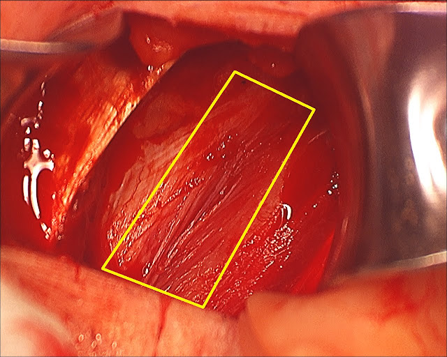
The second photo is another layer down, now looking at the internal oblique. This area wasn't torn, but the area outlined by the yellow box was very thin and barely being held together. It was at risk of tearing at any point. The internal oblique was also torn from the conjoint tendon (but I don't have a photo of that). 
The third photo is showing how Dr. Brown is pulling thicker/stronger portions of the internal oblique together over the thin/compromised area. 
The fourth photo is Dr. Brown pulling the external oblique together (essentially attaching the ends separated by the yellow arrows in photo #1 back together). 
I don't have good photos of the adductor repair or the damaged nerves. Maybe that is a good thing, I don't know if I need to see that as well. This is enough.
|